INSOMNIA DUE TO KNEE PAIN AFTER REPEATED STAIR CLIMBING – WHEN X-RAY AND ULTRASOUND ARE NOT SUFFICIENT
1/30/2026 9:29:21 AM
Background
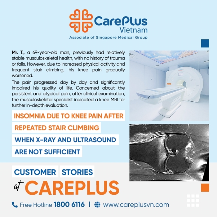
Mr. N.T.M.T., a 69-year-old man, previously had relatively stable musculoskeletal health, with no history of trauma or falls. Due to family-related work demands, over the past two weeks he had to climb stairs and walk more frequently than usual.
Initially, the pain was moderate and improved with rest. However, instead of subsiding, the pain progressively worsened. At its peak, he experienced severe pain even at rest, with nocturnal bone pain that woke him from sleep, significantly impairing his quality of life.
Concerned about the persistent and atypical pain, after clinical examination, the musculoskeletal specialist indicated a knee MRI for further in-depth evaluation.
Knee MRI Findings
The MRI provided a “comprehensive picture” revealing multiple complex lesions that cannot be detected by the naked eye or by basic imaging modalities:
-
Severe osteoarthritis: Predominantly affecting the medial compartment of the knee.
-
Severe meniscal injury:
-
Complex tear of the body and posterior horn of the medial meniscus.
-
Grade 3 tear of the lateral meniscus.
-
Bone marrow lesions (Key finding):
-
Diffuse bone marrow edema of the medial tibial plateau (this is the primary cause of severe nocturnal pain).
-
Patellofemoral joint:
-
Grade 4 cartilage defect (full-thickness cartilage loss) at the lateral facet and central portion of the patella, associated with reactive bone marrow edema.
-
Soft tissue abnormalities:
-
Grade 2 injury of the medial patellar retinaculum.
-
Pes anserine tendinitis.
-
Knee joint effusion and surrounding soft tissue edema.
Clinical interpretation
This case is a typical example of mechanical overload on an aging knee joint. Repeated stair climbing imposed excessive stress on the already weakened menisci and articular cartilage.
As a result, the patient developed complex meniscal tears and full-thickness cartilage loss (grade 4), leading to acute inflammatory reaction and marked bone marrow edema.
It is precisely the bone marrow edema and synovial inflammation that were the main culprits responsible for the severe nocturnal pain that repeatedly awakened the patient at night.
The Value of MRI In Knee Disorders
In this case, MRI demonstrated its role as the optimal imaging modality:
-
Detection of bone marrow edema:
This is the “golden key” of MRI.
-
X-rays only visualize bone structure.
-
Ultrasound only evaluates superficial soft tissues.
-
Only MRI can assess the bone marrow itself and detect early overload-related edema.
-
Accurate evaluation of meniscal tears:
MRI precisely determines the tear pattern and grade (e.g., grade 3, complex tears), guiding the decision on whether arthroscopic meniscal surgery is indicated.
-
Comprehensive assessment:
MRI provides a global view of cartilage, bone, ligaments, menisci, and tendons in a single examination, allowing a thorough explanation of the pain mechanism.
Conclusion
The choice of imaging modality (X-ray, Ultrasound, or MRI) should be guided by a Musculoskeletal Specialist.
Close collaboration between the clinician and the radiologist is the key factor in establishing the most accurate, cost-effective, and efficient treatment strategy for each patient.