THE "HALO" SIGN ON ULTRASOUND – WHEN MRI BECOMES ESSENTIAL TO UNCOVER EARLY LIVER CANCER
8/6/2026 3:12:10 PM
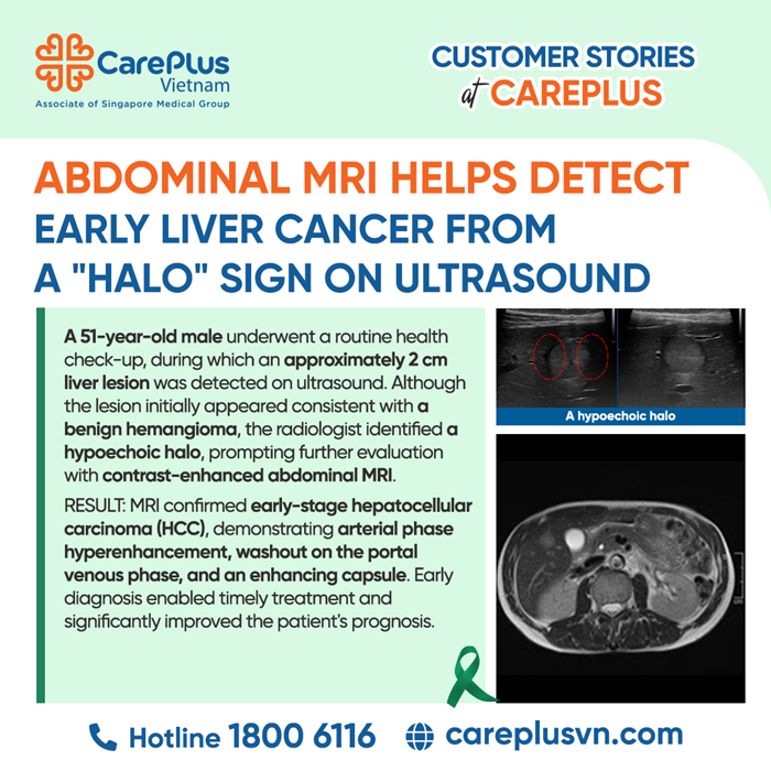
A 51-year-old male with good general health, no history of hepatitis B or C infection, and no alcohol abuse underwent a routine health check-up. During an abdominal ultrasound examination, a 2 cm focal lesion was identified in segment V of the liver.
Morphologically, the lesion appeared hyperechoic with posterior acoustic enhancement, imaging features commonly associated with hepatic hemangioma, one of the most common benign liver tumors. However, the sonographer identified an atypical and concerning finding—a hypoechoic halo surrounding the lesion.
Faced with a lesion demonstrating both benign and suspicious characteristics, the internal medicine physician and radiologist promptly discussed the case. To avoid overlooking a potentially malignant lesion, the patient was immediately referred for a contrast-enhanced abdominal MRI to further characterize the mass.
MRI FINDINGS
The MRI findings completely changed the initial impression of a benign hemangioma and instead demonstrated the classic dynamic enhancement pattern of hepatocellular carcinoma (HCC):
- Arterial phase: Intense and homogeneous arterial hyperenhancement.
- Portal venous and delayed phases: Definite washout, with the lesion becoming hypointense relative to the surrounding liver parenchyma.
- Capsule appearance: A delayed enhancing capsule corresponding to the hypoechoic halo observed on ultrasound.
CLINICAL INSIGHT
This case represents a classic diagnostic pitfall in which early liver cancer could easily have been missed without careful image interpretation.
- The "Low-Risk" Trap
The patient had no history of hepatitis B, hepatitis C, or liver cirrhosis, conditions traditionally associated with a high risk of HCC. Consequently, clinicians may be reassured by the patient's seemingly low-risk profile when an incidental hyperechoic liver lesion is detected.
- The Significance of the Halo
Although a hyperechoic lesion often suggests a benign hemangioma, the presence of a hypoechoic halo is atypical. This finding may represent a pseudocapsule or compressed surrounding liver tissue caused by rapid tumor growth, raising suspicion for malignancy.
- MRI Hemodynamic Features
According to LI-RADS and the AASLD/EASL guidelines, the combination of arterial phase hyperenhancement, portal venous washout, and an enhancing capsule in a liver lesion larger than 1 cm is considered diagnostic of hepatocellular carcinoma.
THE VALUE OF MRI IN CHARACTERIZING FOCAL LIVER LESIONS
This case highlights the complementary roles of ultrasound and MRI in liver imaging.
Ultrasound remains an excellent first-line screening tool because it is widely available, cost-effective, radiation-free, and highly useful for routine health examinations. However, ultrasound has limitations in assessing the vascular behavior of liver lesions.
When ultrasound reveals an indeterminate lesion or suspicious features such as a halo sign, contrast-enhanced MRI becomes the imaging modality of choice. With its superior soft tissue contrast and dynamic multiphase imaging capability, MRI accurately evaluates tumor vascularity and enables confident lesion characterization.
EXPERT MESSAGE: THE RIGHT EXAMINATION LEADS TO THE RIGHT DIAGNOSIS
This case provides an important clinical lesson: a low-risk patient profile should never reduce diagnostic vigilance.
Ultrasound remains the cornerstone of liver screening during routine health check-ups. However, when a liver lesion demonstrates atypical features—such as a halo sign, rapid growth, or abnormal vascular characteristics—contrast-enhanced MRI is strongly recommended to establish an accurate diagnosis.
Close collaboration between clinicians and radiologists enabled this patient's 2 cm hepatocellular carcinoma to be diagnosed at an early stage. Early detection allowed access to potentially curative treatments, including surgical resection or radiofrequency ablation (RFA), significantly improving long-term prognosis and survival.